Back Qolgi metodu Azerbaijani Golgiho metoda Czech Golgi-Färbung German Coloration de Golgi French Golgi-festés Hungarian Metodo di Golgi Italian ゴルジ染色 Japanese Golgikleuring Dutch Metoda Golgiego Polish Метод Гольджи Russian


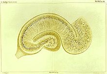
Golgi's method is a silver staining technique that is used to visualize nervous tissue under light microscopy. The method was discovered by Camillo Golgi, an Italian physician and scientist, who published the first picture made with the technique in 1873.[1] It was initially named the black reaction (la reazione nera) by Golgi, but it became better known as the Golgi stain or later, Golgi method.
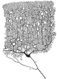
Golgi staining was used by Spanish neuroanatomist Santiago Ramón y Cajal (1852–1934) to discover a number of novel facts about the organization of the nervous system, inspiring the birth of the neuron doctrine. Ultimately, Ramón y Cajal improved the technique by using a method he termed "double impregnation". Ramón y Cajal's staining technique, still in use, is called Cajal's Stain.[citation needed]
- ^ Finger, Stanley (1994). Origins of neuroscience : a history of explorations into brain function. Oxford University Press. p. 45. ISBN 9780195146943. OCLC 27151391.
In 1873, Golgi published the first brief but "adequate" picture of la reazione nera (the black reaction), which showed the whole nerve cell, including its cell body, axon, and branching dendrites.